WEYE
6 Ultrasound-guided system
Specification
Sheet
Main
Technical Parameters
1,
about the system
1.1
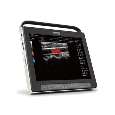
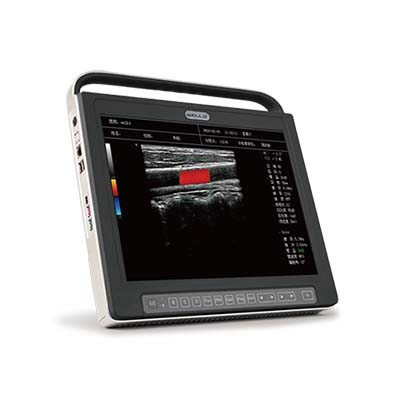
main unit: ultrasound-guided system
An
8-core DSP processor and a front-end ultrasound chip with the
Latest
generation of “digital demodulator” is adopted.
Sparse
transmit & 16 beams parallel processing technology
Pulse
inversion harmonic imaging technology
Synthetic
aperture beam-forming technology
A
continuous transmit focus at every pixel
Speckle
noise suppression technology
Compound
scan technology
1.2
LCD display: ≥ 15 inch touch screen
operation
1.3
Built-in probe connectors: ≥ 2 pcs.
1.4
Display mode: B,BB,B+M,B+C,B+D,B+C+D,PwrD,DirPwr.
1.5
presupposed conditions: preset the check conditions of images optimizing based
on different organs.and these conditions can be freely sorted. Which reduce the
adjustment during operation and the commonly required external regulation and
combined regulation.
1.6 built in lithium battery for continue
working ≥1h
1.7
works on the table, hand held , or wall hanging
1.8
net weight of main unit( exclude battery) ≤3.5kg
Two
- Dimensional gray images
2.1
Linear probe: ≥128 elements, 6.0-13.0 MHz, visible and adjustable
2.2
Gain adjustment range ≥55dB,visible and adjustable.
2.3
Digital zoom: ≥ 10 times pan Zoom in real time
2.4
Image management and record database: built-in integrated ultrasound workshop
with image archiving and record management systems, quick image
storage/retrieve/edit, make fast diagnosis automatically.
2.5
Image storage: one button image storage, built in disk ≥128GB, customized image
storage device available.
2.6
Cine loop: ≥800 frames
2.7
body marks≧100, easy for teaching
2.8
external sports: USB x 2 pcs, Audio, DICOM3.0
2.9
Image Process:Automatic Image Optimization, Enhancement, Smoothing Filter,
Frame Correlation,Sharpen,Pseudo Color,Inversion,Speckle Noise Suppression,
contrast, Line Density,Tissue Harmonic Imaging,etc.
Color/spectra
doppler
3.1
Multi-beam color Ddoppler imaging
3.2
gain range ≥45dB
3.3
color adjustment: Color wall filter, color gain, pulse repetition frequency,
Color
rotary, color sample frame, direction angle etc.
3.4
spectra Doppler adjustment: sample volume,pulse repetition frequency,wall
filter, base line, correction angle etc.
3.5
display area adjustment: linear scanning, target range: -20°to +20°adjustable
Measurement,analysis
and report
4.1
basic measurement: distance,area,volume, angle etc.
4.2
Obstetrics,gynecological measurement,cardiac function measurement ,doppler
Blood
flow measurement and analysis, Measurement and Analysis of peripheral blood
vessels, measurement of small organs, urology and orthopedics ( hips)
measurement and other software package.
4.3
integrated graphic workshop, support DICOM3.0 image transfer,report print,
multi languages.
Standard
configuration
1,color
main unit
2,
high frequency linear probe (128 elements)
3,
Adaptor
4,
power cable
5,
ultrasound gel (250ml)
6,
back support
7,
built in battery
8,
user manual
Optional
configuration
High
frequency linear probe
Transvaginal
probe
Micro
convex probe
Convex
probe
Trolley
cart
Sony
Printer
Working
bag
Battery


