
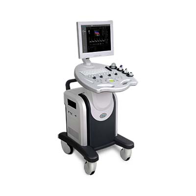
FDC8000 Full Digital Color Doppler Ultrasonic Diagnostic System
- PRODUCT DETAIL
FDC8000
Full Digital Color Doppler Ultrasonic Diagnostic System
Technical Sheet(V1.0)
Device Name:Full Digital Color Doppler
Ultrasonic Diagnostic System
Device Application Use:For
clinic diagnostic needs of Abdomen, Obstetric ,Gynecology,Cardiology, Small
Organs, Superficial Blood Vessels , Musculoskeletal, Anesthesiology,Urology
,neurosurgery , etc.
Characters
1. Global
Leading Ultrasound Platform and Architecture
The latest generation of ultrasonic front-end chip
with "digital demodulator" and 8-core DSP processor provide powerful
computing power, higher integration, lower power consumption as well as clear
image quality.
2. Multi-Beam Parallel Processing Technology
16-beam parallel receiving technology: in B+C mode:abdominal probe depth ≥175mm,
full angle; when ROI frame and B image is coincident, imaging frame rate≥15.0Hz
3. Pulse Inverse Harmonic Imaging (PIHI)
(all probes support Pulse Inverse Harmonic
Technology (PIHI): harmonic operating frequency of High-frequency linear probe
could be 10, 11, 12, 13 MHz four-level adjustable
4. Synthetic Aperture Beam Synthesis Technology(SA)
Totally breaking through the limitation of
traditional DAS beamforming algorithm on the number of physical channels, we
can obtain excellent image quality from near field to far field with smaller
hardware scale and lower transmitting power
5. Emission Point-by-Point Focusing Technology
(Full-range Focusing)
No focus position display ,no need to adjust the
focus manually
6.Speckle noise suppression technology
“mclear”,Four levels adjustable;
Effectively remove the speckle noise in ultrasound
images to obtain a clearer and more delicate 2D image
7.Free-hand 3D +4D imaging
For fetal examination ,greatly improve the
ultrasound detection radio of abnormal fetuses
Three active probe sockets, freely switch on
,support hot-plug
DICOM3.0 ,With network remote assistance tool
software, technicians can maintain the system remotely through the network
Technical Parameters:
1 System
general parameters:
1.1 Imaging
Mode:B、2B、4B、B+M、B+C、real
time B+C+D、B+D、PwrD、DirPwr
1.2
Input/Output port:USB2.0,DICOM3.0,VGA,RS-232
1.3 Optional
Probe:
Convex probe(R60/3.5MHz/128-element)
High Frequency Linear probe(L38/7.5MHz/128-element)
Endo-cavity (R10/6.5MHz/128-element)
Micro-convex(R20/3.5MHz/128-element)
4D Volume probe(R40/3.5MHz/128-element)
1.4
Measurement
It has a variety of measurement packages for
Abdomen,Obstetrics and Gynecology,Urology,Cardiology,Orthopaedics,Superficial
,etc.
Using scalar map measurement markers, a measurement
item can be deleted,moved,dragged and adjusted arbitrarily,and the measurement
results will be changed dynamically
1.5
Annotation: Bod mark and text notes; Body Marks ≥100
1.6 Image
storage: Image can be saved in JPG format ,which can browse multiple images in
the same interface
1.7 Cine loop: Cine Loop ≥1000 frames,which support
playback single frame, manual/automatic playback, and can capture a single
image during playback.The formats includes AVI and can be reloaded, replayed
and measured
1.8 Files
Management
a、Report: Support case management,
graphic and text integration workstation, report editing, preview, printing
b、SSD≥128G, Supports one-key rapid
access to medical records, support external connecting CD-ROM
recording/erasing, U-disk data backup.
c、Has the work list module connected
with PACS server, the hospital PACS system can be accessed through LAN to
realize DICOM 3.0 image network management.
2.B mode
2.1 Beam
Former: Full Digital Beam Former
2.2 Received
Beam Focusing: Continuous Dynamics
2.3 TGC:≥8
seg
2.4 Tissue
Harmonic:2-level adjustable. pulse inversion harmonic in 2D mode and
conventional fundamental wave in other modes
2.5 Enlarge
Functionality: Zoom ≥ 10 times
2.6 Scanning
depth: continuously adjustable
2.7 Scanning
angle:continuously adjustable
2.8 Gray
scale:256
2.9 Scanning
lines ≥25
2.10 Pseudo
:10
2.11 Gain:
can be adjusted independently in real time in B/M
2.12 Image processing: 8-level frame correlation,
5-level line correlation, image post-processing, line density, image
up/down/left flip, image rotation (2B, 4B mode 180 degrees rotation);
3. M mode
3.1 Scanning speed: four level adjustable
3.2 Gain can be adjusted independently in real time
3.3 The Position of Sampling line adjustable
4.B+C mode
4.1 Size ,postion and color of Sample Volume
adjustable,Can support the width of sample volume is same in B/C
4.2 PRF:
Convex :0.5~5.0 kHz grade adjustable
Linear:0.5-5.0 kHz grade
adjustable
4.3
Deflection Angle: The range of the orientation angle of the image of
interest in linear array scanning can be adjusted by grading from -15°~
+15°
4.4 Wall
Fliter :10 grade adjustable
4.5 Color
Gain:21 dB ~45dB
4.6 Color
priority: 20-250 continuous adjustable
4.7 Color
afterglow: 7levels adjustable
4.8 Color reversal
and automatic tracing
5. B+D Mode
5.1 Pulse
Repetition Frequency:
Convex : 0.5-1.25kHz ,4 levels adjustable(Open
Update),0.5-7.5kHz
24 levels adjustable(Close Update)
Linear:0.5-3.0kHz 11 levels
adjustable(Open update)、0.5-7.5kHz 24levels adjustable(Close
Update)
5.2 Gain:
19dB ~ 48dB, grading adjustable
5.3 Wall
Filter:
Convex : 10-125 Hz , adjustable(Open
Update),40-500Hz adjustable(Close Update)
Linear:30-375Hz adjustable(Open
Update)、150-1875Hz adjustable(close update)
5.4 Noise
reduction: 01dB-32dB
5.5 Sample
Volume:0.5-20mm adjustable
5.6 Sweep
Speed:8 levels adjustable
5.7 Support
for synchronous updates, automatic tracing and spectrum inversion
5.8 Baseline
,Spectrum sound volume and Correction angle adjustable
5.9 Supporting the above, below and all tracing of
baseline, the tracing can be smoothed and the parameters of the smoothing can
be adjusted. Can realize the real-time calculation of heart rate, maximum speed
and average speed
5.10 Blood
Flow measured Speed:
a) Maximum measured blood flow velocity in Pulse
Doppler status ≥8 m/s
b) Minimum measured blood flow velocity:≤2 mm/s
(non-noise signal)
Software
1. Design with upgrade ability can meet the need of
expanding clinical application.
The software is the latest version and contains all
the functions that have been released.
Compliance with Standards
1. GB10152-2009 B-mode Ultrasound Diagnostic
Equipment
2. YY 0767-2009 Ultrasound Color Flow Imaging
System
3. YY 0505-2012 Medical Electrical Equipment Part
1-2: General Safety Requirements Parallel to Standard EMC Requirements and
Tests
4. GB9706.1-2007 Medical Electrical Equipment Part
1: General Safety Requirements
5. GB9706.9-2008 Medical Electrical Equipment Part
2-37: Special Requirements for the Safety of Ultrasound Diagnostic and
Monitoring Equipment
Configuration
Standard Configuration
Main unit with three active probe connectors and
15” LCD (1 pcs)
Convex probe (R60/3.5MHz/128-element,1pcs
)
High frequency Linear probe (L38/7.5MHz/128-element
,1pcs)
Power cable (1.8m)
Gel(250ml)
Fuse(2A,250V)
Optional Configuration
Transvaginal (R10/6.5MHz/128-element)
Micro-convex (R20/3.5MHz/128-element)
4DVolume probe +4D software (R40/3.5MHz/128-element)
Foot Switch
Printer
Biopsy Bracket
19 inches LCD
Main unit with four active probe connectors


