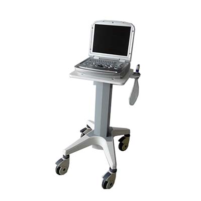
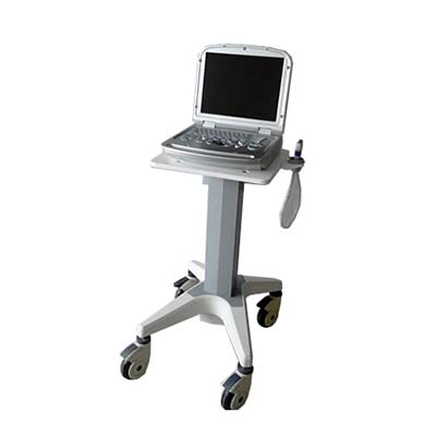
MDK-9600MDK Full
Digital Color Doppler Ultrasound Diagnostic System
Introduction:
Equipment
use: Abdomen, blood vessels, breast, obstetrics and gynecology, superficial
structures, musculoskeletal, urology, pediatrics/newborns, small organs, heart,
etc.
Technical Parameters:
1.
System general functions
1.1
Monitor: 15-inch high-resolution color LCD monitor, no flicker, can be up,
down, left, and right
Spin.
1.2
Operation keyboard: Silicone button, flexible and convenient operation.
1.3
Probe interface ≥1, fully activated, interoperability
2.
Probe specifications
2.1
Probe frequency: ultra-wideband probe, imaging frequency range 2.0MHz—12.00MHz
2.2
Both two-dimensional and Doppler (B/D): linear array B/PWD; convex array B/PWD
2.3
Puncture guide: optional probe can be equipped with puncture guide device
(optional)
3.
The main parameters of two-dimensional imaging:
3.1
Scan
Abdominal
convex array: frequency 2.0-5.0MHz, five-segment frequency conversion
Superficial
linear array: frequency 6.0-12.0MHz, five-segment frequency conversion
Vaginal
probe: frequency 5.0-10MHZ, five-segment frequency conversion
Phased
array probe: frequency 2.0-5.0 five-segment frequency conversion
3.2
Maximum display depth: ≥30cm
3.3
Real-time ZOOM zoom function
3.4
Sound beam deflection of linear array probe: ≥10°
3.5
Sound speed focus: ≥4 focus points, position continuously adjustable
3.6
Playback reproduction: large-capacity movie playback
3.7
Ultrasonic output power: visually adjustable
3.8
Digital sound beam former: continuous dynamic focus, variable aperture and
dynamic zoom
3.9
Preset conditions: Preset the inspection conditions for the optimized image for
different inspection organs, reducing the adjustment during operation
*3.10
Image optimization technology: visually adjustable ≥6 files
3.11
Gain adjustment: 8-segment TGC adjustment
3.12
M scan speed ≥ 3 gears adjustable
3.13
B/M layout is visually adjustable
4.
Pulse Doppler
4.1
Deflection angle: -2.5-10 adjustable in four stages
4.2
Pulse repetition frequency: 1.82KHZ-7.52KHZ adjustable
4.3
Spectrum flip
4.4
Triple synchronization
4.5
Wall filter ≥ 3 levels adjustable
4.6
Spectrum pseudo color ≥6 adjustable
4.7
Spectrum envelope function: multiple modes of real-time spectrum envelope are
optional, and the system automatically analyzes and displays: PSV, EDV, AT, DT,
RI, PI, SD, HR and other data
4.8
Edge enhancement ≥ 8 kinds of adjustable
4.7
Baseline≥7 adjustable
5.
Color Doppler
5.1 Doppler probe and frequency: linear
array: PWD; convex array: PWD
All
of the above probes have 2 or more Doppler imaging operating frequencies and
independently adjustable
5.2
Wall filtering: ≥3 levels, continuously adjustable
5.3 Display position adjustment: the image
range of interest for linear scanning: -10°~ +10°
6. Ultrasound image and medical record
management system
6.1 The compression ratio of dynamic images
can be adjusted, and it can be directly installed on a normal PC without
special software.


